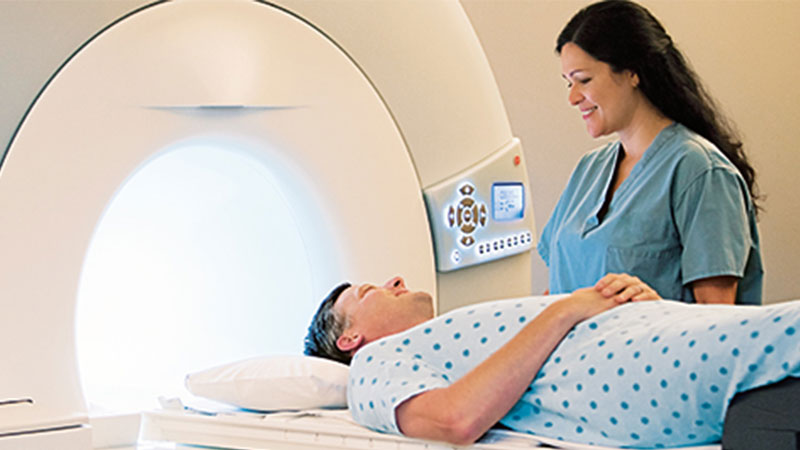
A Note On echocardiogram
An echocardiogram (reverberation) is a realistic plan of your heart’s development. During a reverberation test, your healthcare provider uses ultrasound (high-recurrence sound waves) from a handheld wand placed on your chest to take pictures of your heart’s valves and chambers. This helps the provider assess your heart’s siphon activity. Vendors often combine reverberation with Doppler ultrasound and various Doppler methods to assess blood flow in the heart valves. Echocardiography does not use radiation. This makes echocardiogram in Sparta, NJ not the same as different tests, such as X-rays and CT scans, which use modest amounts of radiation.
Who does a reverb test?
A specialist called a cardiovascular sonographer reproduces its reverberation. They are prepared to perform reverb tests and utilize the latest innovation. They are ready to work in different environments, including medical clinic rooms and catheterization labs.
What procedures are used in echocardiography?
Some strategies can be used to make pictures of your heart. The best strategy depends on your particular condition and what your supplier needs to see. These methods incorporate
Two-layer ultrasound (2D). This approach is used most often. Produces 2D images that appear as “slices” on the PC screen. These cuts can often be “stacked” to build a 3D design.
Three-layer ultrasound (3D). Progress in innovation has made 3D imaging more productive and useful. New 3D methods show various parts of your heart, including how well it sucks blood, with more remarkable accuracy. Using 3D also allows your sonographer to see parts of your heart from various points.
Doppler ultrasound. This method shows you how fast your blood flow and, in addition, in which direction.
Variety Doppler ultrasound. This method also shows your bloodstream but uses several varieties to show the various bearings of the flow.
Stress images. This approach shows changes in the way the heart muscle moves. You can get early indications of some coronary heart disease.
Contrast image. Your supplier infuses a substance called a differentiation specialist into one of your veins. The substance is noticeable in the photos and can help to show the subtleties of your heart. Certain individuals experience an unfavorably susceptible response to the differentiation specialist, but the responses are usually mild.